Articoli correlati a Atlas of Endoscopic Anatomy for Endonasal Intracranial...

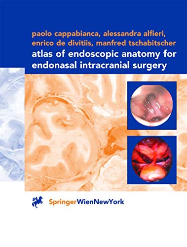
It is only recently that the use of the endoscope as the sole visualizing tool has been introduced in transsphenoidal pituitary surgery with its favorable related implications and minimal operative trauma. Of course, microscopic and endoscopic anatomy are basically the same, but the optical distorsion of endoscopic images is quite substantial compared to microscopic depictions. An endoscope lens produces images with maximal magnification at its center and severe contraction at its periphery. Nearer images are disproportionally enlarged and remote images are falsely miniaturized. This optical illusion may disorientate a surgeon who is not familiar with this peculiar condition at the skull base. This atlas acts as a guide through the endoscopic anatomy and gives detailed descriptions of the preoperative management and the surgical procedures.
Le informazioni nella sezione "Riassunto" possono far riferimento a edizioni diverse di questo titolo.
Recensione:
"... Den Autoren ist ein vom ausgezeichneten Bildmaterial dominierter Atlas gelungen ... " Annals of Anatomy 185/5/2003
Contenuti:
I. Anatomic preparations.- I.A. Gross anatomy.- I.A.1. Bone preparations.- I.A.2. Nose and paranasal sinuses.- I.B. Endoscopic surgical anatomy.- I.B.1. Nose.- I.B.2. Sphenoidal sinus.- I.B.3. Sella turcica region.- I.B.4. Suprasellar region.- I.B.5. Parasellar region.- I.B.6. Retrosellar region.- II. Preoperative management.- II.A. Neuroradiological investigations.- II.A.1. CT.- II.A.2. MRI.- II.B. Operating theatre.- II.B.1. Positioning of the patient.- II.B.2. Equipment.- III. Surgical procedure.- III.A. Surgical steps.- III.A.1. Endonasal approach to the sphenoidal sinus ostium.- III.A.2. Enlargement of the sphenoidal sinus ostium.- III.A.3. Preparation of the sphenoid sinus.- III.A.4. Opening of the floor of the sella turcica.- III.A.5. Opening of the dura mater.- III.A.6. Removal of the lesion.- III.A.7. Sella turcica reconstruction.- Appendix: Selected clinical cases.- Case 1: Intra-suprasellar macroadenoma.- Case 2: Intra-parasellar macroadenoma.- Case 3: Solid intra-suprasellar craniopharyngeoma.- Case 4: Cystic intra-suprasellar craniopharyngeoma.- Case 5: Arachnoid intra-suprasellar cyst.- Case 6: Intra-suprasellar RATHKE’s cleft cyst.- References.
Le informazioni nella sezione "Su questo libro" possono far riferimento a edizioni diverse di questo titolo.
- EditoreSpringer Verlag Wien
- Data di pubblicazione2001
- ISBN 10 3211835482
- ISBN 13 9783211835487
- RilegaturaCopertina rigida
- Numero di pagine150
Compra nuovo
Scopri di più su questo articolo
EUR 129,65
Spese di spedizione:
EUR 5,11
In U.S.A.
I migliori risultati di ricerca su AbeBooks
ATLAS OF ENDOSCOPIC ANATOMY FOR
Da:
Valutazione libreria
Descrizione libro Condizione: New. New. In shrink wrap. Looks like an interesting title! 1.9. Codice articolo Q-3211835482
Compra nuovo
EUR 129,65
Convertire valuta

