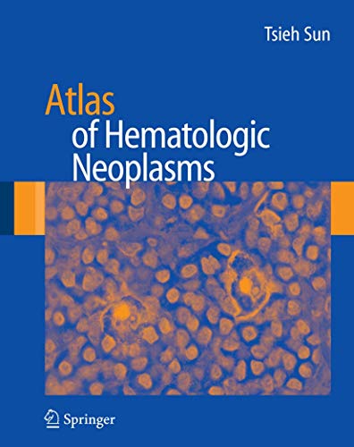
Articoli correlati a Atlas of Hematologic Neoplasms

Due to its rapid development in recent years, hematopathology has become a very complicated discipline. The current development is mainly in two aspects: the new classification of lymphomas and leukemias and the new techniques.
The Revised European-American Classification of Lymphoid Neoplasms (REAL classification) and the World Health Organization (WHO) classification of hematologic neoplasms require not only morphologic criteria but also immunophenotyping and molecular genetics for the diagnosis of hematologic tumors. Immunophenotyping is performed by either flow cytometry or immunohistochemistry. There are many new monoclonal antibodies and new equipments accumulated in recent years that make immunophenotyping more or more accurate and helpful. There are even more new techniques invented in recent years in the field of molecular genetics. In cytogenetics, the conventional karyotype is supplemented and partly replaced by the fluorescence in situ hybridization (FISH) technique. The current development of gene expression profiling is even more powerful in terms of subtyping the hematologic tumors, which may help guiding the treatment and predict the prognosis. In molecular biology, the tedious Southern blotting technique is largely replaced by polymerase chain reaction (PCR). The recent development in reverse-transcriptase PCR and quantitative PCR makes these techniques even more versatile.
Because of these new developments, hematopathology has become too complicated to handle by a general pathologist. Many hospitals have to hire a newly trained hematopathologist to oversee peripheral blood, bone marrow and lymph node examinations. These young hematopathologists are geared to the new techniques, but most of them are inexperienced in morphology. No matter how well-trained a hematopathologist is, he or she still needs to see enough cases so that they can recognize the morphology and use the new techniques to substantiate the diagnosis. In other words, morphology is still the basis for the diagnosis of lymphomas and leukemias.
Therefore, a good color atlas is the most helpful tool for these young hematopathologists and for the surgical pathologists who may encounter a few cases of hematologic tumors from time to time. In a busy daily practice, it is difficult to refer to a comprehensive hematologic textbook all the time. There are a few hematologic color atlases on the market to show the morphology of the normal blood cells and hematologic tumor cells. These books are helpful but not enough, because tumor cell morphology is variable from case to case and different kinds of tumor cells may look alike and need to be differentiated by other parameters.
The best way to learn morphology is through the format of clinical case study. This format is also consistent with the daily practice of hematopathologists and with the pattern in all the specialty board examinations. Therefore, it is a good learning tool for the pathology residents, hematology fellows as well as medical students.
This proposed book will present 83 clinical cases with clinical history, morphology of the original specimen and a list of differential diagnoses. This is followed by further testing with pictures to show the test results. At the end, a correct diagnosis is rendered with subsequent brief discussion on how the diagnosis is achieved. A few useful references will be cited and a table will be provided for differential diagnosis in some cases.
The major emphasis is the provision of 500 color photos of peripheral blood smears, bone marrow aspirates, core biopsy, lymph node biopsy and biopsies of other solid organs that are involved with lymphomas and leukemias. Pictures of other diagnostic parameters, such as flow cytometric histograms, immunohistochemical stains, cytogenetic karyotypes, fluorescence in situ hybridization and polymerase chain reaction, will also be included.
A comprehensive approach with consideration of clinical, morphologic, immunophenotypic and molecular genetic aspects is the best way to achieve a correct diagnosis. After reading this book, the reader will learn to make a diagnosis not only based on the morphology alone but also in conjunction with other parameters.
Le informazioni nella sezione "Riassunto" possono far riferimento a edizioni diverse di questo titolo.
From the reviews:
"Exactly as the title states, this is an atlas of hematologic neoplasms. ... Intended for ‘young’ hematopathologists and surgical pathologists, this atlas also would be of great interest to laboratory medicine and/or pathology residents in training, hematopathology fellows, clinical hematology-oncology fellows, and their counterparts in practice. ... This is a very useful atlas to have ready at hand to assist with hematologic disorder diagnosis. I highly recommend it and I also recommend you chain it to your bookcase ... ." (Valerie L Ng, Doody’s Review Service, November, 2009)
“This atlas does a fine job of placing the morphologic diagnosis on centre stage, between the clinical findings and the subsequent confirmatory tests. ... The atlas contains more than 500 high-resolution photographs ... . Atlas of Hematologic Neoplasms can be read for enjoyment one case at a time as individual clinicopathological conferences. Alternatively, the cases can be studied for board reviews and teaching sessions. The book will definitely be helpful for students, trainees, or experienced clinicians in general pathology, hematopathology, hematology, or oncology ... .” (Paulette Mehta, Journal of the American Medical Association, Vol. 303 (6), 2010)Hematopathology has become a very complicated discipline. New classification of lymphomas and leukemias as well as new techniques are rapidly moving the field forward. Atlas of Hematologic Neoplasms, written by Tsieh Sun, MD, is designed with general pathologists, pathologists-in-training, clinical hematologists, and oncologists in mind. It presents over eighty clinical cases with clinical history, morphology of the original specimen, and differential diagnoses.
A concise discussion, references and teaching tables are available for further exploration. The major asset of this book is the provision of more than 500 (or 541, to be exact) beautiful photos depicting the morphology of peripheral blood smear, bone marrow, lymph node and other organs, as well as flow cytometric and molecular genetic pictures to illustrate the diagnostic principles.
Le informazioni nella sezione "Su questo libro" possono far riferimento a edizioni diverse di questo titolo.
- EditoreSpringer Verlag
- Data di pubblicazione2009
- ISBN 10 0387898476
- ISBN 13 9780387898476
- RilegaturaCopertina rigida
- Numero edizione1
- Numero di pagine525
- RedattoreSun Tsieh M.D.
Compra nuovo
Scopri di più su questo articolo
Spese di spedizione:
EUR 3,29
In U.S.A.
I migliori risultati di ricerca su AbeBooks
Atlas of Hematologic Neoplasms
Descrizione libro Hardcover. Condizione: new. New. Codice articolo Wizard0387898476
Atlas of Hematologic Neoplasms
Descrizione libro Hardcover. Condizione: new. Buy for Great customer experience. Codice articolo GoldenDragon0387898476
Atlas of Hematologic Neoplasms
Descrizione libro Hardcover. Condizione: new. New Copy. Customer Service Guaranteed. Codice articolo think0387898476
Atlas of Hematologic Neoplasms
Descrizione libro Condizione: new. Codice articolo FrontCover0387898476
Atlas of Hematologic Neoplasms
Descrizione libro Hardcover. Condizione: new. New. Fast Shipping and good customer service. Codice articolo Holz_New_0387898476
Atlas of Hematologic Neoplasms
Descrizione libro Gebunden. Condizione: New. Dieser Artikel ist ein Print on Demand Artikel und wird nach Ihrer Bestellung fuer Sie gedruckt. Quick referenceDifferential diagnosis based learningWritten for the general pathologist or pathologist-in-trainingCompletely illustrated in colorDue to its rapid development in recent years, hematopathology has become a ve. Codice articolo 5911559
Atlas of Hematologic Neoplasms
Descrizione libro Condizione: New. Codice articolo 6543981-n
Atlas of Hematologic Neoplasms
Descrizione libro Buch. Condizione: Neu. This item is printed on demand - it takes 3-4 days longer - Neuware -Due to its rapid development in recent years, hematopathology has become a very complicated discipline. The current development is mainly in two aspects: the new classification of lymphomas and leukemias and the new techniques.The Revised European-American Classification of Lymphoid Neoplasms (REAL classification) and the World Health Organization (WHO) classification of hematologic neoplasms require not only morphologic criteria but also immunophenotyping and molecular genetics for the diagnosis of hematologic tumors. Immunophenotyping is performed by either flow cytometry or immunohistochemistry. There are many new monoclonal antibodies and new equipments accumulated in recent years that make immunophenotyping more or more accurate and helpful. There are even more new techniques invented in recent years in the field of molecular genetics. In cytogenetics, the conventional karyotype is supplemented and partly replaced by the fluorescence in situ hybridization (FISH) technique. The current development of gene expression profiling is even more powerful in terms of subtyping the hematologic tumors, which may help guiding the treatment and predict the prognosis. In molecular biology, the tedious Southern blotting technique is largely replaced by polymerase chain reaction (PCR). The recent development in reverse-transcriptase PCR and quantitative PCR makes these techniques even more versatile. Because of these new developments, hematopathology has become too complicated to handle by a general pathologist. Many hospitals have to hire a newly trained hematopathologist to oversee peripheral blood, bone marrow and lymph node examinations. These young hematopathologists are geared to the new techniques, but most of them are inexperienced in morphology. No matter how well-trained a hematopathologist is, he or she still needs to see enough cases so that they can recognize the morphology and use the new techniques to substantiate the diagnosis. In other words, morphology is still the basis for the diagnosis of lymphomas and leukemias.Therefore, a good color atlas is the most helpful tool for these young hematopathologists and for the surgical pathologists who may encounter a few cases of hematologic tumors from time to time. In a busy daily practice, it is difficult to refer to a comprehensive hematologic textbook all the time. There are a few hematologic color atlases on the market to show the morphology of the normal blood cells and hematologic tumor cells. These books are helpful but not enough, because tumor cell morphology is variable from case to case and different kinds of tumor cells may look alike and need to be differentiated by other parameters. The best way to learn morphology is through the format of clinical case study. This format is also consistent with the daily practice of hematopathologists and with the pattern in all the specialty board examinations. Therefore, it is a good learning tool for the pathology residents, hematology fellows as well as medical students. This proposed book will present 83 clinical cases with clinical history, morphology of the original specimen and a list of differential diagnoses. This is followed by further testing with pictures to show the test results. At the end, a correct diagnosis is rendered with subsequent brief discussion on how the diagnosis is achieved. A few useful references will be cited and a table will be provided for differential diagnosis in some cases. The major emphasis is the provision of 500 color photos of peripheral blood smears, bone marrow aspirates, core biopsy, lymph node biopsy and biopsies of other solid organs that are involved with lymphomas and leukemias. Pictures of other diagnostic parameters, such as flow cytometric histograms, immunohistochemical stains, cytogenetic karyotypes, fluorescence in situ hybridization and polymerase chain reaction, will also be included.A comprehensive approach with cons 540 pp. Englisch. Codice articolo 9780387898476
Atlas of Hematologic Neoplasms
Descrizione libro Buch. Condizione: Neu. Druck auf Anfrage Neuware - Printed after ordering - Due to its rapid development in recent years, hematopathology has become a very complicated discipline. The current development is mainly in two aspects: the new classification of lymphomas and leukemias and the new techniques.The Revised European-American Classification of Lymphoid Neoplasms (REAL classification) and the World Health Organization (WHO) classification of hematologic neoplasms require not only morphologic criteria but also immunophenotyping and molecular genetics for the diagnosis of hematologic tumors. Immunophenotyping is performed by either flow cytometry or immunohistochemistry. There are many new monoclonal antibodies and new equipments accumulated in recent years that make immunophenotyping more or more accurate and helpful. There are even more new techniques invented in recent years in the field of molecular genetics. In cytogenetics, the conventional karyotype is supplemented and partly replaced by the fluorescence in situ hybridization (FISH) technique. The current development of gene expression profiling is even more powerful in terms of subtyping the hematologic tumors, which may help guiding the treatment and predict the prognosis. In molecular biology, the tedious Southern blotting technique is largely replaced by polymerase chain reaction (PCR). The recent development in reverse-transcriptase PCR and quantitative PCR makes these techniques even more versatile. Because of these new developments, hematopathology has become too complicated to handle by a general pathologist. Many hospitals have to hire a newly trained hematopathologist to oversee peripheral blood, bone marrow and lymph node examinations. These young hematopathologists are geared to the new techniques, but most of them are inexperienced in morphology. No matter how well-trained a hematopathologist is, he or she still needs to see enough cases so that they can recognize the morphology and use the new techniques to substantiate the diagnosis. In other words, morphology is still the basis for the diagnosis of lymphomas and leukemias.Therefore, a good color atlas is the most helpful tool for these young hematopathologists and for the surgical pathologists who may encounter a few cases of hematologic tumors from time to time. In a busy daily practice, it is difficult to refer to a comprehensive hematologic textbook all the time. There are a few hematologic color atlases on the market to show the morphology of the normal blood cells and hematologic tumor cells. These books are helpful but not enough, because tumor cell morphology is variable from case to case and different kinds of tumor cells may look alike and need to be differentiated by other parameters. The best way to learn morphology is through the format of clinical case study. This format is also consistent with the daily practice of hematopathologists and with the pattern in all the specialty board examinations. Therefore, it is a good learning tool for the pathology residents, hematology fellows as well as medical students. This proposed book will present 83 clinical cases with clinical history, morphology of the original specimen and a list of differential diagnoses. This is followed by further testing with pictures to show the test results. At the end, a correct diagnosis is rendered with subsequent brief discussion on how the diagnosis is achieved. A few useful references will be cited and a table will be provided for differential diagnosis in some cases. The major emphasis is the provision of 500 color photos of peripheral blood smears, bone marrow aspirates, core biopsy, lymph node biopsy and biopsies of other solid organs that are involved with lymphomas and leukemias. Pictures of other diagnostic parameters, such as flow cytometric histograms, immunohistochemical stains, cytogenetic karyotypes, fluorescence in situ hybridization and polymerase chain reaction, will also be included.A comprehensive approach with cons. Codice articolo 9780387898476

