
Importo totale (1 articolo articoli):
Destinazione ordine:

demian e s (20 risultati)
Vai alla pagina principale dei risultati di ricerca
Filtri di ricerca
Tipo di articolo
- Tutti i tipi di prodotto
- Libri (20)
- Riviste e Giornali (Nessun altro risultato corrispondente a questo perfezionamento)
- Fumetti (Nessun altro risultato corrispondente a questo perfezionamento)
- Spartiti (Nessun altro risultato corrispondente a questo perfezionamento)
- Arte, Stampe e Poster (Nessun altro risultato corrispondente a questo perfezionamento)
- Fotografie (Nessun altro risultato corrispondente a questo perfezionamento)
- Mappe (Nessun altro risultato corrispondente a questo perfezionamento)
- Manoscritti e Collezionismo cartaceo (Nessun altro risultato corrispondente a questo perfezionamento)
Condizioni Maggiori informazioni
- Nuovo (2)
- Come nuovo, Ottimo o Quasi ottimo (Nessun altro risultato corrispondente a questo perfezionamento)
- Molto buono o Buono (Nessun altro risultato corrispondente a questo perfezionamento)
- Discreto o Mediocre (Nessun altro risultato corrispondente a questo perfezionamento)
- Come descritto (18)
Legatura
- Tutte
- Rilegato (Nessun altro risultato corrispondente a questo perfezionamento)
- Brossura (2)
Ulteriori caratteristiche
- Prima ed. (Nessun altro risultato corrispondente a questo perfezionamento)
- Copia autograf. (Nessun altro risultato corrispondente a questo perfezionamento)
- Sovracoperta (Nessun altro risultato corrispondente a questo perfezionamento)
- Con foto (14)
- Non Print on Demand (20)
Lingua (2)
Prezzo
- Qualsiasi prezzo
- Inferiore a EUR 20
- EUR 20 a EUR 45
- Superiore a EUR 45 (Nessun altro risultato corrispondente a questo perfezionamento)
Spedizione gratuita
- Spedizione gratuita in U.S.A. (Nessun altro risultato corrispondente a questo perfezionamento)
Paese del venditore
Valutazione venditore
-

El rostro oculto del animal
Da: KALAMO BOOKS, Burriana, CS, Spagna
Valutazione del venditore 5 su 5 stelle

EUR 11,50
Spedizione EUR 17,86
Spedito da Spagna a U.S.A.Quantit�: 1 disponibili
Aggiungi al carrelloTapa blanda. Condizione: Nuevo.
-

ANTOLOG�A PO�?TICA. Manías del corazón
Lingua: Spagnolo
Editore: GRUPO EDITORIAL LETRAS NEGRAS S.A.S
ISBN 10: 6287680156 ISBN 13: 9786287680159
Da: KALAMO BOOKS, Burriana, CS, Spagna
Valutazione del venditore 5 su 5 stelle
EUR 13,50
Spedizione EUR 17,86
Spedito da Spagna a U.S.A.Quantit�: 1 disponibili
Aggiungi al carrelloTapa blanda. Condizione: Nuevo.
-

The egg mass, egg laying and mating behaviour of the snail Marisa cornuarietis (L.)
Editore: 1970-1971, 1970
EUR 5,75
Spedizione EUR 24,00
Spedito da Germania a U.S.A.Quantit�: 1 disponibili
Aggiungi al carrello12 pp., 3 figs, gr. 8.
-
EUR 5,75
Spedizione EUR 24,00
Spedito da Germania a U.S.A.Quantit�: 1 disponibili
Aggiungi al carrello12 pp., 5 figs, gr. 8.
-

Prospects of the use of Marisa cornuarietis in the biological control of Limnaea cailliaudi in th U.A.R
Data di pubblicazione: 1965
EUR 5,75
Spedizione EUR 24,00
Spedito da Germania a U.S.A.Quantit�: 1 disponibili
Aggiungi al carrello5 pp., 3 pls, gr. 8.
-

Predatory activity of Marisa cornuarietis against Bulinus (Bulinus) truncatus, the transmitter of urinary schistosomiasis
Data di pubblicazione: 1965
EUR 5,75
Spedizione EUR 24,00
Spedito da Germania a U.S.A.Quantit�: 1 disponibili
Aggiungi al carrello6 pp. + 4 pls, gr. 8.
-
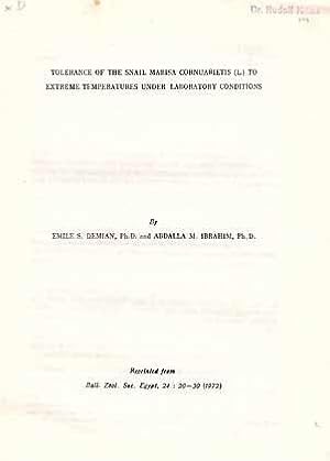
Tolerance of the snail Marisa cornuarietis (L.) to extreme temperature under laboratory conditions
Data di pubblicazione: 1972
EUR 5,75
Spedizione EUR 24,00
Spedito da Germania a U.S.A.Quantit�: 1 disponibili
Aggiungi al carrello11 pp., 4 figs, gr. 8.
-

Embryonic development and organogenesis in the snail Marisa cornuarietis (Mesogastropoda: Ampullariidae). V. Development of the nervous system
Data di pubblicazione: 1975
EUR 5,75
Spedizione EUR 24,00
Spedito da Germania a U.S.A.Quantit�: 1 disponibili
Aggiungi al carrelloThe nervous system is ectodermal in origin. All nerve ganglia arise separately by proliferation and later delamination from the ectoderm, not by invagination. They become secondarily connected to one another by commissures and connectives developing as extensions from the peripheral layer of ganglionic nerve cells. Rudiments of the cerebral, pedal, pleural and intestinal (parietal) ganglia arise almost simultaneously at a relatively early stage (Stage V). The cerebral ganglia develop from the ectoderm of the head plates. Rudiments of the pedal and pleural ganglia are separate at their inception. They later fuse (Stage VI) to form a pleuro-pedal ganglionic mass on each side. The 2 intestinal ganglia are symmetrical at the beginning, but they soon lose their symmetry as a result of torsion. The right ganglion crosses to the left over the gut and persists as the supraintestinal ganglion. The left or subintestinal ganglion shifts to the right and forward, and fuses with the right pleural ganglion (Stage VIII), thus obscuring the chiastoneury. The paired buccal and single visceral (abdominal) ganglia start differentiating in Stage VII. The former develop from the ectodermal wall of the stomodaeum, while the visceral ganglion delaminates from the right wall of the visceral sac, then shifts to the left during torsion. The statocysts develop early (Stage V) from 2 ectodermal invaginations on either side of the rudimentary foot. They later separate from the overlying ectoderm and statoconi appear in their lumina. Contrary to earlier reports on related ampullariids, the osphradium proved to be ontogenetically older than the mantle and mantle cavity. It starts differentiating as a thickened ectodermal plate in the right wall of the visceral sac (Stage V). During torsion, it becomes engulfed in the mantle cavity and shifts to the left side, then is carried forward as the mantlegrow. The eyes develop late (Stage IX) as ectodermal invaginations which rapidly separate from the ectoderm to form closed vesicles. Their cells start differentiating before hatching to form the retina, in which pigment is deposited, and the inner cornea. The lens is secreted in the lumen of the eye and grows by addition of concentric layers of secretion. 14 pp., 10 figs, gr. 8.
-

Studies on the Chromosome Numbers in the Ampulariidae (Gastropoda, Prosobranchia)
Data di pubblicazione: 1965
EUR 5,75
Spedizione EUR 24,00
Spedito da Germania a U.S.A.Quantit�: 1 disponibili
Aggiungi al carrelloTreated taxa: Marisa cornuarietis, Lanistes bolteni, Pila ovata 6 pp. + 3 pls, 4.
-

Embryonic development and organogenesis in the snail Marisa cornuarietis (Mesogastropoda: Ampullariidae). IV. Development of the shell gland, mantle and respiratory organs
Data di pubblicazione: 1973
EUR 5,87
Spedizione EUR 24,00
Spedito da Germania a U.S.A.Quantit�: 1 disponibili
Aggiungi al carrello17 pp., 13 figs, gr. 8.
-

Embryonic development and organogenesis in the snail Marisa cornuarietis (Mesogastropoda: Ampullariidae). III. Development of the circulatory and renal system
Data di pubblicazione: 1973
EUR 6,90
Spedizione EUR 24,00
Spedito da Germania a U.S.A.Quantit�: 1 disponibili
Aggiungi al carrelloThe nervous system is ectodermal in origin. All nerve ganglia arise separately by proliferation and later delamination from the ectoderm, not by invagination. They become secondarily connected to one another by commissures and connectives developing as extensions from the peripheral layer of ganglionic nerve cells. Rudiments of the cerebral, pedal, pleural and intestinal (parietal) ganglia arise almost simultaneously at a relatively early stage (Stage V). The cerebral ganglia develop from the ectoderm of the head plates. Rudiments of the pedal and pleural ganglia are separate at their inception. They later fuse (Stage VI) to form a pleuro-pedal ganglionic mass on each side. The 2 intestinal ganglia are symmetrical at the beginning, but they soon lose their symmetry as a result of torsion. The right ganglion crosses to the left over the gut and persists as the supraintestinal ganglion. The left or subintestinal ganglion shifts to the right and forward, and fuses with the right pleural ganglion (Stage VIII), thus obscuring the chiastoneury. The paired buccal and single visceral (abdominal) ganglia start differentiating in Stage VII. The former develop from the ectodermal wall of the stomodaeum, while the visceral ganglion delaminates from the right wall of the visceral sac, then shifts to the left during torsion. The statocysts develop early (Stage V) from 2 ectodermal invaginations on either side of the rudimentary foot. They later separate from the overlying ectoderm and statoconi appear in their lumina. Contrary to earlier reports on related ampullariids, the osphradium proved to be ontogenetically older than the mantle and mantle cavity. It starts differentiating as a thickened ectodermal plate in the right wall of the visceral sac (Stage V). During torsion, it becomes engulfed in the mantle cavity and shifts to the left side, then is carried forward as the mantlegrow. The eyes develop late (Stage IX) as ectodermal invaginations which rapidly separate from the ectoderm to form closed vesicles. Their cells start differentiating before hatching to form the retina, in which pigment is deposited, and the inner cornea. The lens is secreted in the lumen of the eye and grows by addition of concentric layers of secretion. 20 pp., 10 figs, gr. 8.
-
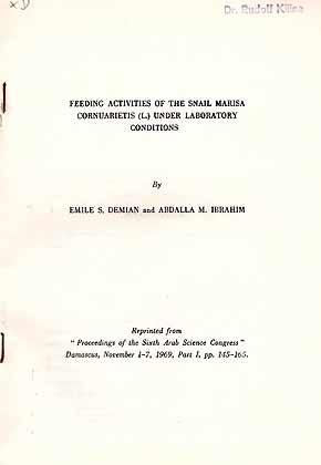
Feeding activities of the snail Marisa cornuarietis (L.) under laboratory conditions
Data di pubblicazione: 1969
EUR 7,25
Spedizione EUR 24,00
Spedito da Germania a U.S.A.Quantit�: 1 disponibili
Aggiungi al carrello21 pp., 2 figs, gr. 8.
-

EUR 7,25
Spedizione EUR 24,00
Spedito da Germania a U.S.A.Quantit�: 1 disponibili
Aggiungi al carrello21 pp., 9 figs, stapled gr. 8.
-

Embryonic development and organogenesis in the snail Marisa cornuarietis (Mesogastropoda: Ampullariidae). II. Development of the alimentary system
Data di pubblicazione: 1973
EUR 8,28
Spedizione EUR 24,00
Spedito da Germania a U.S.A.Quantit�: 1 disponibili
Aggiungi al carrello24 pp., 16 figs, gr. 8.
-
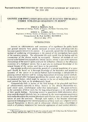
Growth and population dynamics of Bulinus truncatus under semi-field conditions in Egypt
Data di pubblicazione: 1972
EUR 8,28
Spedizione EUR 24,00
Spedito da Germania a U.S.A.Quantit�: 1 disponibili
Aggiungi al carrello24 pp., 13 figs, gr. 8.
-
Prospects of the use of Marisa cornuarietis in the biological control of Limnaea caillaudi in the U.A.R. In 4�, offp., pp. 8 with 3 pls. Offprint from Proc. Egyptian Ac. Sci., 18
Data di pubblicazione: 1965
Da: Riccardo Giannuzzi Savelli, Palermo, PA, Italia
Valutazione del venditore 2 su 5 stelle

EUR 3,50
Spedizione EUR 30,00
Spedito da Italia a U.S.A.Quantit�: 1 disponibili
Aggiungi al carrello -

Embryonic development and organogenesis in the snail Marisa cornuarietis (Mesogastropoda: Ampullariidae). I. General outlines of development
Data di pubblicazione: 1973
EUR 9,66
Spedizione EUR 24,00
Spedito da Germania a U.S.A.Quantit�: 1 disponibili
Aggiungi al carrello28 pp., 24 figs, gr. 8.
-

The histology of the alimentary system of Marisa cornuarietis (Mesogastropoda: Ampullariidae)
Data di pubblicazione: 1967
EUR 9,89
Spedizione EUR 24,00
Spedito da Germania a U.S.A.Quantit�: 1 disponibili
Aggiungi al carrello48 pp., 59 figs, gr. 8.
-
Embryonic development and organogenesis in the snail Marisa cornuarietis (Mesogastropoda: Ampullariidae) - II Development of the alimentary system. In 8vo, offp., pp. 24 with 16 figs. Offprint from Malacologia 12(1)
Data di pubblicazione: 1973
Da: Riccardo Giannuzzi Savelli, Palermo, PA, Italia
Valutazione del venditore 2 su 5 stelle
EUR 4,00
Spedizione EUR 30,00
Spedito da Italia a U.S.A.Quantit�: 1 disponibili
Aggiungi al carrello -
Collection of 17 papers on anatomy, physiology and development of Marisa cornuarietis.
Editore: 1964-1975, 1964
Da: Hermann L. Strack, Loguivy Plougras, Francia
Valutazione del venditore 5 su 5 stelle
EUR 38,50
Spedizione EUR 48,00
Spedito da Francia a U.S.A.Quantit�: 1 disponibili
Aggiungi al carrelloAll are stapled or in printed wrappers. Some quite large like ??Anatomy of the alimentary system of marisa cornuarietis?? (75 p., 21 figs). All ex library Dr. A.C. van Bruggen (with his stamp).
